Electro-encephalography is the study of the electrical activity of the brain. It is based on the fact that neurons, the grey cells of the brain, communicate with each other by sending electrical signals known as action potentials and post-synaptic potentials. While the action potential is not perceptible on the surface, the post-synaptic potential, comparable to a dipole, can be recorded. The direction of the neurons also has an important effect on the distribution of the electric field. Only pyramidal cells possess a special orientation and contribute to the surface potential. Their arrangement in perpendicular macro-columns on the surface of the cerebral cortex is one of the reasons why it is possible to record their output. The electroencephalogram (EEG) records the electrical activity incorporated in a large number of neurons, each electrode receiving a signal from hundreds of thousands of cells.
There are two types of recording, surface and penetrating. Surface recording is performed using a cap containing twenty or so electrodes arranged symmetrically, making it possible to cover the whole of the cerebral cortex. This non-invasive examination is routinely performed in an out-patient setting. Penetrating recording is performed using electrodes arranged in a grid pattern on the actual surface of the brain, or using penetrating electrodes inserted into the brain. This requires surgery, and is only performed in hospital in response to very specific indications.
An EEG is the only examination that directly measures electrical activity in the brain in real time. Its temporal resolution, of the order of a few milliseconds, makes it possible to perform a detailed analysis of how this activity develops and is extremely useful, for example, for analyzing the propagation of epileptic seizures.
The indications involved are very extensive:
-
epileptic seizures of all types (tonic-clonic, absence, partial, complex or simple, i.e. with or without loss of contact, etc.)
-
fainting or sudden loss of consciousness
-
states of confusion
-
neurological paroxystic phenomena (déjà-vu/déjà-vécu, for example).
-
variations in states of consciousness, of severity up to coma
-
metabolic disorders (electrolytic problems, such as sodium deficiency, liver or renal failure, etc.)
-
infections of the brain (encephalitis)
-
Creutzfeldt-Jakob disease
-
insomnia
-
dementia or the suspicion of cognitive disturbance.
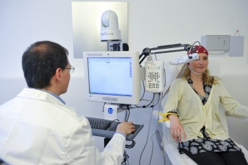
The standard examination performed in an out-patient environment involves the recording of the surface of the brain, and lasts for about 20 minutes. A total of about 45 minutes is required, including preparation time. The patient is seated comfortably in an examination armchair with the cap arranged on his/her head. To reduce resistance to a minimum, as this could alter the signal between the scalp and the electrodes in the cap, a gel is applied to the scalp at the point of each electrode. Two electrodes or probes are applied to the wrists, in order to measure the heart rate throughout the examination.
Several manœuvres are performed during the procedure in order to study the basic brain waves and highlight any anomalies. The patient is asked to open and close his/her eyes several times, which makes it possible to study the basic rhythm. The patient is then asked to perform a hyperpnea, requiring him/her to breathe slowly and deeply for three minutes. This first exercise induces hypocapnia (a reduction in the concentration of CO2 in the blood) leading to alkalosis, causing the blood vessels in the brain to constrict. The brain is thus subjected to pressure through a reduction in its oxygenation. The object is to detect a change in the basic rhythm, an accentuation of pre-existing anomalies or the detection thereof. The test can be performed on patients from the age of three; the main contra-indications are severe heart or respiratory disorders. Once basic oxygenation has been restored to the brain, which will take a few minutes, a second provocation test is performed, consisting of intermittent light stimulation. The patient is exposed to a series of light flashes; his/her eyes are closed at first, then open, for various lengths of time. The purpose is to detect abnormal activity that is light-dependent (photo-paroxystic). The patient is then made to feel as calm as possible for the last part of the recording, even allowing him/her to doze, since numerous anomalies can be revealed in the early stages of sleep. It is often useful, especially in cases of strong clinical suspicion, to repeat the test to increase sensitivity.
Following this standard surface recording of brain activity, a recording may be undertaken during a short sleep (mainly for children) or under sleep deprivation conditions (mainly for adults). This test, performed using the same cap, lasts longer (up to 90 minutes of recording time) and is designed to explore brain activity in the first stages of sleep. Furthermore, depriving the patient of sleep enhances the sensitivity of the examination, and increases the chances of detecting anomalies. The patient lies comfortably on the examination couch and, after being asked to open and close his/her eyes several times, he/she is allowed to sleep for about one hour.
Finally, lengthy surface recordings may be performed, in a hospital setting only. In this case, electrodes are individually adhered to the scalp and the patient’s brain activity is recording for a minimum of one night.
All these examinations are performed in tandem with video recording, making it possible to correlate electro-encephalographic anomalies and any associated clinical symptoms. In some cases, this analysis can also make it possible to exclude a neurological origin for certain suspect symptoms if the patient experiences these symptoms during the recording, although no abnormality is observed in the EEG.